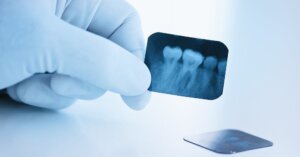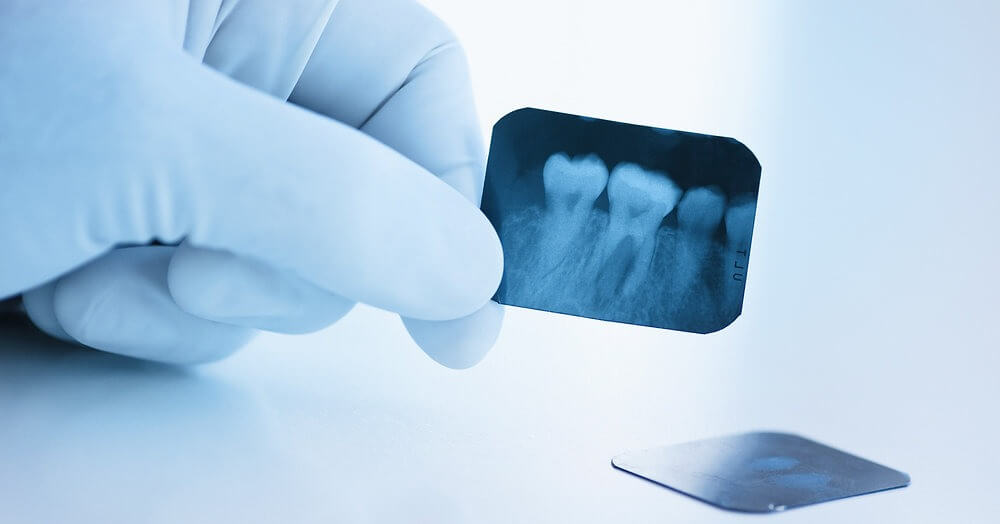
Dental x-rays (radiographs) provide valuable information not only about your teeth but also about your jaw bone, nerves and sinuses. We use digital X-rays that emit up to 90% less radiation than traditional film X-rays.
First, your hygienist will cover you with a lead apron or thyroid collar to minimize radiation exposure. Then the X-ray sensor or film is placed inside your mouth or, in the case of panoramic X-rays, around your head.
Benefits
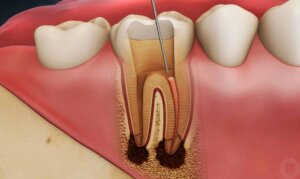
Dental x-ray tampa fl are valuable diagnostic tools that help dentists see hidden areas of decay between your teeth and below existing fillings. X-rays can also reveal abscessed or impacted teeth, gum disease, cysts and tumors. Early detection saves you time, money and discomfort.
Digital x-rays are more comfortable for patients and expose up to 90% less radiation than traditional film x-rays. Using a digital sensor instead of a piece of film, the x-ray image appears instantly on a computer screen. The images can be enhanced or magnified for better diagnosis. They can also be sent electronically to specialists or insurance companies.
An x-ray of your jaws shows the status of your wisdom teeth and can detect jaw problems, such as temporomandibular joint (TMJ) issues. Panoramic x-rays provide a wider view of the entire mouth area and can detect impacted or unerupted teeth, bone infections or abnormalities, cysts, tumors and sinuses.
X-rays can also show some types of oral cancer, but they cannot detect all forms of mouth cancer. To be fully screened, you should schedule regular visits to your dentist. If you have no dental insurance, a financing plan may be available for you to spread out the cost of your care. CareCredit is an example of a popular option. AARP also offers dental benefits for seniors. You can also look into the many options for supplemental health insurance.
Types
X-rays use electromagnetic radiation to detect hidden structures like your teeth and jawbone. These images allow your dentist to see underlying problems before they can be seen by the naked eye, such as tooth decay, bone loss and cysts or abscesses. Dental X-rays are available in traditional (using film) and digital formats (using sensors and computers). Digital dental X-rays require less exposure to radiation than their traditional counterparts.
There are several different types of dental x-ray tampa fl, including bitewing, periapical and panoramic. Bitewing X-rays show the entire tooth, its roots and the surrounding bone. They are usually taken at recall (checkup) visits to detect new cavities between your teeth and check for bone loss. Periapical X-rays capture more information about the teeth and their roots, such as the degree of bone loss, impacted or unerupted teeth and jaw fractures. Pediatric dentists frequently use occlusal X-rays to examine developing teeth.
Panoramic X-rays provide a wraparound view of your upper and lower teeth, the nasal area, sinuses and temporomandibular joints (TMJ). These X-rays are commonly used to detect impacted wisdom teeth, monitor jaw growth and development, evaluate bone loss and diagnose cysts or abscesses. Panorex X-rays are extraoral and easy to perform. The sensor or film is placed outside of the mouth and a mechanism rotates around the head to take pictures from multiple angles.
Safety
In the past, X-rays were feared for their radiation exposure but, since then, techniques and equipment have been honed to minimise this. The amount of radiation used for a dental X-ray is very small. The American Academy of Oral and Maxillofacial Radiology explains that the amount of external radiation scatter (which also affects the rest of your body) from a single digital X-ray is equivalent to the amount of radiation absorbed by your body during one aeroplane flight across the country. In addition, the number of X-rays taken is comparable to the amount of background radiation you get daily from the sun, earth, soil and rocks.
Having a digital X-ray takes less time than traditional film-based X-rays, and emits up to 90% less radiation. You can trust that your dentist and their team will only use the minimum amount of radiation required to obtain a high-quality image.
The coordinator of the radiation protection program has responsibilities to support facility owners, dentists, dental hygienists, oral surgeons and dental assistants with regard to procedures, radiography equipment performance and radiation safety. They perform tasks such as conducting facility radiation surveys, assessing the need for personal dosimeters and calculating facility shielding requirements. The coordinator may also be responsible for performing quality assurance testing of X-ray equipment. This includes visual inspection and X-raying protective devices to ensure they are not defective, or that their structural integrity is intact.
Comfort
X-rays use electromagnetic radiation to capture internal images of your mouth and jawbone. The process is quick, easy and painless. In fact, digital X-rays emit up to 90% less radiation than traditional film X-rays.
Our dental team uses state-of-the-art technology for your safety and comfort. The images from your X-rays are transmitted digitally to our computer. This allows your dentist to examine them instantly. Moreover, the digital images can be enlarged, enhanced or sharpened for easier diagnoses of oral conditions.
The images are very accurate, enabling your dentist to spot the smallest issues. This way, he can catch problems in their earliest stages and prevent them from progressing to more serious dental and medical problems. Besides, X-rays can reveal tooth decay, impacted wisdom teeth, cysts, tumors, bone fractures and other issues that cannot be spotted with just visual examination.
Depending on your medical history and symptoms, your dentist may also recommend other imaging tests. Some of these include:

